Blogs
Beyond the Basics: A Layman's Guide to the Slit Lamp Microscope

You have likely seen the intimidating machine with lights and mirrors that you sit in front of during an eye test with a doctor. The doctor puts their ear to the wall, tinkers with several knobs, and looks into a small telescope at your face. It is a slit lamp microscope, one of the most significant tools used in eye care.
The little slit lamp may look elaborate, but it is a genius yet simple idea that assists doctors in examining the eye with fine detail. So, here is a more straightforward explanation of this fantastic piece of medical equipment.
What Exactly Is a Slit Lamp Microscope?
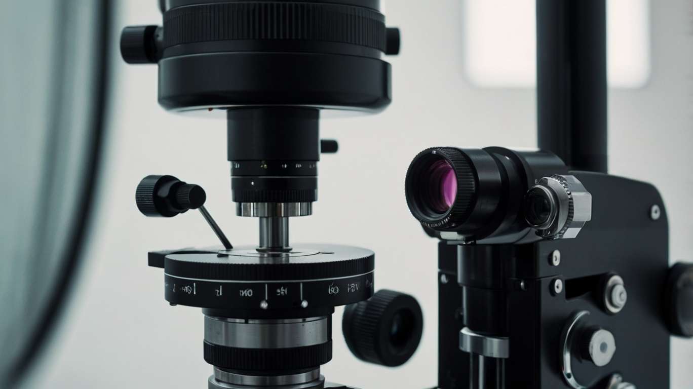
Consider a slit lamp as a high-powered magnifying glass and a very accurate, pointed flashlight. The slit component is the result of the small beam of light produced by it, which can be made narrow by a small line or widened into a thicker beam. The lamp provides the light, and the microscope section magnifies everything, allowing the doctor to see small details.
The entire system is placed on a table, and both you and the doctor occupy specific positions. The doctor looks through the microscope section, and you put your chin on a rest, staring straight ahead. They are designed to provide the doctor with a clear view of your eyes while ensuring your comfort and well-being.
It does the magic as the light beam falls on your eye at an angle, as the doctor's observation is made through the microscope at an angle. This arrangement provides a three-dimensional image of your eye structures, which would otherwise be impossible to observe.
The Two Main Parts: Light and Magnification
The Illumination System
The light system is more similar to a super-precise flashlight, which can be regulated in numerous ways. A bright, clean, white light is provided by a bright halogen or LED bulb, usually used as a light source. Some filters and lenses reflect this light and end up in your eye.
It is an adjustable slit. How about peeping through a keyhole compared to peeking through a door? The slit functions just like that. A thin beam can cut through various layers in your eye, and a wide slit lets in a wide range of light.
The doctor is also able to adjust the position of the light, making it appear in various directions. This is particularly important since certain eye complications manifest only when light is directed to a specific side of the eye.
The Observation System
The microscope itself is a highly advanced pair of binoculars attached to an adjustable stand. It also gives a magnification of approximately 6-40 times the standard size, depending on the model and the settings.
The number of different magnification levels that the doctor can adjust on most slit lamps is extensive. Lower magnification shows the entire eye area, while high magnification allows the doctor to focus on specific details. Designed as binoculars, it provides depth. The three-dimensional picture helps physicians determine the magnitude of another structure or how deeply issues may extend within the eye.
The Process of Examination:
The first time you sit down for a slit lamp exam, it may seem like a mysterious process, but it is pretty simple. The physician begins by aligning the equipment at your height level and eye position.
Doctors examine your eyes, including your eyelashes and eyelids, as well as the cornea. They may request that you blink or look in various directions to indicate how everything moves.
Different Types of Light Techniques
There are several lighting methods applied in the examination by doctors, and each technique provides different information:
· Direct illumination is similar to placing a flashlight on an object. This highlights surface-level information and apparent issues.
· Indirect illumination is reflected against the structures around to produce subliminal effects of light. This method makes visible issues that are concealed in direct light.
· Retro illumination features light that emanates from around the structures and glows. This is ideal for examining clear areas of the eye and detecting cloudiness, as well as any abnormalities.
What Can Doctors View Using Such an Incredible Tool?
The slit lamp provides a fantastic level of detail about your eyes. Doctors are capable of examining all major structures, both internally and externally.
The Front of the Eye:
The eyelids and the eyelashes may appear to be a simple thing, but they can contain numerous issues. The slit lamp can detect minute mites, oil glands blocked, or indications of inflammation that the naked eye cannot detect.
The clear portion of your eye (called the sclera) is closely scrutinized, and the white one is also scrutinized, as well as the clear covering above the colored portion of your eye (conjunctiva). The color (redness) and swelling or abnormal development are clear under a microscope.
The Cornea:
The slit lamp excels most significantly in examining the cornea. This thick, dome-like anterior part of your eye is only half a millimeter thick, though the slit lamp may scan through this in layers.
Doctors detect scratches, infections, and cloudy areas that blur vision. They measure cornea thickness and check for signs of swelling, which may indicate potential problems.
In the Anterior Chamber:
The back of the cornea contains clear fluid known as the anterior chamber. A slit lamp can examine this space to determine whether it is inflamed, bleeding, or otherwise.
Your eye color (the iris) and the black hole in the center of the eye (the pupil) are closely checked as well. Discolorations of the iris or abnormal shapes of the pupil may indicate any eye disease.
Why This Tool Is So Important?
The slit lamp microscope is a marvel that has revolutionized eye care, as it enables doctors to detect any problem that would have remained unseen until it became severe. Most eye diseases begin with minor alterations that are not noticeable to the naked eye in normal lighting conditions.
Many eye diseases should be identified early. Another example is glaucoma, which can gradually rob you of sight, with no signs until the damage is in an advanced stage. The slit lamp can help diagnose or detect the onset of glaucoma even before you are aware of it.
Special Techniques and Add-ons
Contemporary slit lamps are capable of much more than simple analysis. Most of them have special attachments and methods that vastly increase their capabilities.
Pressure Measurement
Certain slit lamps can even measure pressure in your eye (by applanation tonometry). A tiny probe is softly placed on the front of your eye (after numbing drops) to gauge the amount of pressure required to distribute the inside of a highly tiny region. The test plays a critical role in the identification of glaucoma.
Documentation and Photography
Many contemporary slit lamps are equipped with cameras, which can capture images of what the physician sees up close. Such images can be stored in your medical file, sent to other physicians, or used to track changes over time.
Fluorescein Staining
Physicians sometimes use fluorescein, an orange dye, to highlight specific eye issues. It shows scratches, dry areas, or injuries on the eye surface and appears bright green under the slit lamp's blue light.
The Human Touch in High-Tech Medicine
Despite all its high technology, slit lamp still needs human talent to become truly useful. It takes years for doctors to become trained in the use of all the various lighting techniques, as well as to understand what they observe.
It is an art to know which technique to apply and when, how to vary the lighting according to the patient, and to identify specific nuances that may suggest serious complications. A computer cannot substitute a trained eye and the experience of an expert eye care specialist.
Final Words
This slit lamp microscope may appear to be a beast, but it is your weapon on the path to healthy eyes. This is a highly advanced piece of equipment that enables physicians to examine the eyes in great detail, detect early defects, and monitor treatment progress.
The next time you are in front of one, you will be aware that you are using one of the most advanced and notable instruments of modern medicine.
