Blogs
Choosing the Right Fundus Camera: Mydriatic vs. Non-Mydriatic and Their Applications
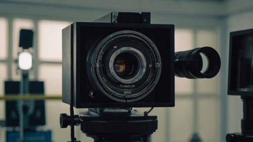
Fundus cameras are like the backstage passes of eye care. If your eyeballs are throwing a tantrum because of diabetes, glaucoma, macular degeneration, or even just high blood pressure, these little machines are the ones catching all the action. They capture these amazingly sharp images of the back of your eye. This way, your eye doctor can track changes, spotting the issues early, and keeping your vision in check. Fundus cameras are not one-size-fits-all. Some of them, such as mydriatic ones, require dilation. But the non-mydriatic cameras? No drops, just snap and go. That one little difference —dilate or don’t—decides how these cameras are used in the clinical environment. Some doctors prefer the traditional approach, while others opt for a quicker and easier method. Either way, these cameras are the unsung heroes, ensuring your eyes stay out of trouble. What is a Mydriatic Fundus Camera? Traditional mydriatic fundus cameras require the instillation of mydriatic eye drops to dilate the pupil before image acquisition. Clinicians dilate the pupil to have a wide, clear view of the retina, especially in the peripheral regions. This technique reliably delivers the best images because the camera can focus and operate at its level of excellence. A full range of applications makes these units the best choice when combining wide-field imaging, fluorescein angiography (FA), indocyanine green angiography (ICG), or mydriatics in general. However, dilation is not without cost; applying the drops may take time, typically requiring 15 to 30 minutes before any effect is noticeable. Patients may suffer ill effects during that time. Brightness and blurriness are also effects that can persist for several hours after the examination, which can be inconvenient for patients who plan to return to work or drive. This can also be a problem for those with narrow-angle glaucoma and those allergic to the use of eye drops. Nonetheless, mydriatic fundus cameras would remain the best in their class under the gold standard set, especially in specialized situations such as those typically found in ophthalmology clinics, where deep-seated retinal pathology needs to be well-documented and closely followed. What is A Non-Mydriatic Fundus Camera? In contrast, non-mydriatic fundus cameras feature a special design that enables the capture of a retina image even when the pupil is not dilated. These cameras capture images in the infrared frequency and are equipped with sophisticated optics, enabling them to map and image pupils as small as 3.5 mm in diameter. These cameras are widely used in various eyecare settings, including primary care clinics, diabetic retinopathy programs, and outreach programs. All waiting time associated with dilation and patient discomfort is minimized with non-mydriatic systems. The exam can be completed within a few minutes, making it ideal for high-throughput situations and when rapid assessment is needed. The patient can resume several activities immediately after the test, promoting compliance and overall satisfaction. Nevertheless, the usual disadvantage of a non-mydriatic system is the narrow field of view, which is often limited to 30 to 45 degrees, and is not able to adequately document peripheral retinal pathology as well as mydriatic systems do. Additionally, image quality may be compromised with small pupils, or in some patients with media opacities resulting from cataracts or vitreous hemorrhage. Clinical Workflow and Patient Experience Aspect Mydriatic Fundus Camera Non-Mydriatic Fundus Camera Patient Preparation Requires pharmacologic dilation (15–30 minutes wait time) No dilation needed; ready for imaging immediately. Appointment Duration Longer due to dilation and recovery time. Shorter, typically completed in a few minutes. Patient Comfort Causes include temporary discomfort, light sensitivity, and blurry vision as after-effects. Minimal discomfort and does not result in visual disruption after the examination session. Post-Exam Activity Patients may need assistance with transportation and daily tasks. Patients can resume normal activities immediately. Staff Involvement Additional staff may be needed to administer and monitor dilation Fewer staff resources required; simpler operation Workflow Efficiency A slower pace for in-depth exams. High productivity is suited for screening and routine documentation. Setting Suitability Ophthalmology clinics and retinal specialists will find them particularly effective. Perfect fit for primary care, community clinics, and telemedicine. Patient Compliance May be lower due to inconvenience and recovery time Higher compliance due to a fast, comfortable experience. Applications in Diabetic Retinopathy and Telemedicine Aspect Mydriatic Fundus Camera Non-Mydriatic Fundus Camera Use in Diabetic Retinopathy (DR) Utilized to document advanced cases requiring detailed imaging and angiography. The best application is for routine DR screenings and early detection. Imaging Capabilities Fluorescein angiography (FA) and wide-field imaging are supported. Can only deliver standard central retinal imaging. Telemedicine Suitability A bad choice due to size, cost, and need for dilation Works well in telemedicine since it is portable, compact, and easily integrated with telehealth. Deployment in Remote Areas Not easy as the equipment has a complex design and dilation requirement. Works well in rural environments. Speed of Image Capture Slower; dilation is required before imaging. Fast - enables rapid image capture. Patient Population Focus Best for referred patients with complex or sight-threatening conditions. Ideal for screening large populations in primary care or diabetes clinics. Follow-up and Referral Use Best for confirmation diagnosis and treatment guidance. Used for triage, which involves positive findings referred for further evaluation. Cost Considerations and Practice Integration Aspect Mydriatic Fundus Camera Non-Mydriatic Fundus Camera Initial Equipment Cost A high price tag, as it includes advanced imaging features and specialized functions. More affordable, suited for clinics with limited budgets. Operational Complexity Requires experienced personnel for dilation and imaging User-friendly, and only basic training is required. Space Requirements Bulky size that requires a dedicated imaging room. Compact and portable, it can fit in a smaller clinical space. Maintenance and Upkeep Maintenance costs are higher due to the more complex optics and accessories. Lower maintenance as it contains fewer moving parts and the design is simple. Practice Type Compatibility Best fit for specialty ophthalmology and retina-focused practice. Versatile and compatible with all types of practices: primary care, optometry, and general practice settings Scalability Less scalable in high-volume or mobile settings. Highly scalable; suitable for outreach programs and telehealth expansion. Return on Investment (ROI) Long-term ROI through advanced diagnostics and treatment planning. Faster ROI in screening programs as patient turnover is lower and the overhead is lower. Making the Right Choice for Your Practice Choosing a fundus camera isn’t like picking out any ordinary gadget. It’s more about understanding the clinical setting and the nature of your work. You’ve gotta vibe with your workflow, know your patients, and keep your clinical goals in mind. Got a waiting room packed with folks needing diabetes screenings? Non-mydriatic is the perfect instrument for a speedy diagnosis: quick snaps, no dilating, excellent patient satisfaction. But suppose you’re the type who loves a good close-up, preferring to see every millimeter of retinal detail. In that case, there's nothing better than a mydriatic fundus camera with its big, bold, crystal-clear images—perfect for those high-stakes treatment choices. No matter what, though, you’re always playing a balancing act: efficiency vs. image quality vs. patient comfort vs. what your inner perfectionist demands. Once you get a feel for these cameras’ quirks and superpowers, you’ll be serving up top-notch care and, let’s face it, feeling pretty slick about your gear choices.
Mastering the Slit Lamp Microscope: A Step-by-Step Guide for Beginners
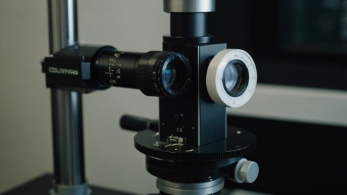
Using a slit lamp may be a frustrating experience to learn. All eye doctors have this complicated-looking instrument, and you, as an inexperienced learner, may tend to ask yourself how in the world you will ever come to grips with all the moving levers and buttons? It takes the proper technique and practice to use a slit lamp comfortably. The slit lamp is your window to the eye. It is like a super-powered magnifying lens and a pinpoint light source that enables one to see what the human eye could never sense. As soon as you grasp the fundamentals, you will find out that it is a very simple tool that uses rational steps. What is Slit Lamp Microscope? So, what are we supposedly working with before we discuss how to work with it? A slit lamp essentially is a combination of both a microscope and a light source. The light creates a narrow slit (that would be the slit bit) which you can took to shine on various sections of the eye. The microscope component enables magnification of the observed object. Imagine this way, suppose you were to examine a clear glass marble in a dark room. Well, you would want a flashlight to see through it, wouldn't you? The slit lamp does the same with eyes, with an extensive increase in precision and strength. Preparing: Your Pre-Exam Checklist 1. Creating a Work Area Ask your patient to sit down before being prepared. Ensure that the slit lamp is clean, the power is connected, and all controls move smoothly. A test of the light and magnification takes time afterwards. Have tissues available, in case patients become emotional during the examination. Make sure your instruments are nearby--fluorescein drops, numbing drops, or tonometry equipment that you will use. 2. Preparing Yourself Find the seat height that feels comfortable to you. Keep your back straight and arms resting naturally. Wash your hands and be mentally prepared to concentrate. A slit lamp examination is sensitive, so it is essential to remain undistracted during the examination. Patient Positioning: Making It Right in the Beginning 3. Initial Setup Assist your patient into the chair and adjust it to a height where their eyes are level with the instrument. It may require several attempts; each patient is unique, and such precision with height will result in a higher quality of the examination. Request that the patient sit straight with their feet flat on the floor. Their good posture also assists them when taking the exam. When they are slouching or feel uneasy, they will move more, and that will cause you to work more. 4. The Chin Rest and Forehead Bar Direct the patient's chin to the chin rest. This should not be uncomfortable, though. Their foreheads only need to be resting on the forehead bar, not squeezed. One helpful tip is to instruct the patients to rest their foreheads on the bar, not pressing it. Too much pressure is exerted by many people, which causes tension and increases their chances of relocating. 5. Eye Level Adjustment The patient's eyes must be level with the instrument's reference marks. Adjust the table up or down instead of having the patient crane their neck or slouch if it is too high or too low. Ensure that the eyes are aligned straight. Patients may unknowingly lean their head, an aspect that can disrupt your examination. Instrument Adjustment: How to Make It Work Your Way? 6. Beginning with the Basics The magnification starts at 10x. This will provide you with a broader perspective and help you orient more easily. One thing that you can always do later is magnify to view details. Adjust the level of lighting to an acceptable level. Excessive brightness can stun the patient and may cause tears to be shed. Too much darkness makes it impossible to notice details. Begin by using a medium and adding it as required. 7. The Slit Beam settings Begin with slit beam using medium width which is kept vertical. This will be your normal initial point of all examinations. The beam must be of a level that can sufficiently light a structure, but not to a level of discomfort. The depth of the slit must be big enough to cover the area you wish to investigate. To suit general viewing, make it tall so that it lights up the entire visible portion of the eye. 8. Targeting the Instrument Operate the whole set of instruments by working the joystick. Minor manipulations are critical, as excessive jerkiness can cause uneasiness for both the examiner and the patient. Concentration by forth and back sliding of the whole slit lamp itself, and not by separately moving parts. This puts everything in place. Simple Techniques of Examination 9. To begin with, the Lids and Lashes You must always start by examining the eyelids and eyelashes. This will enable you to practice with the controls as you examine familiar objects, such as easily visible structures. 10. To the Conjunctiva The conjunctiva is the transparent layer on the white surface of the eye. It is a good second act since it is relatively simple to observe and test. Have the patient look in various directions to examine different areas of the body. 11. The Cornea Examination Slit lamp examination is of real value in the cornea. It is a visible portion of the eye that may conceal the invisible dissimilarities. An angle of a narrow-slit beam will then be used to form what can be termed as an optical section. This process illuminates the cornea as a slice, making issues within the tissue layers more visible. 12. The Anterior Chamber The area between the cornea and iris is called the anterior chamber. It should be full of clear fluid; however, issues in this area may indicate severe eye diseases. Check whether it is clear, there are floating particles, or if there are depth changes. The slit beam method is also helpful in this case, as the light is directed at an angle to create contrast between normal and abnormal tissue. Tips on Success 1. Communication is Priority Communicate with your patients during the check-up. Inform them about your business and what they can expect from it. If a patient is blinking excessively, allow them to rest. A compelling study seldom achieves good results. 2. Beginner Mistakes • Do not maximize your magnification. You might want to zoom in now, but it could cause you to lose your orientation and miss the big picture. • Make sure not to hurry between the various areas. It is beneficial to take your time to examine each structure closely before proceeding. • Make a point to check both eyes. One becomes so focused on checking one eye that one forgets to examine the other to make a comparison. 3. Developing Your Skills Master the simple controls when you are not on schedule. Learning how to focus, shift the slit beam, and adjust magnification significantly facilitates real examinations. Observe good professionals as often as possible. Pay attention to the postures of patients, settings, and their way of passing during their examination routine. Fixing Common Issues 1. When You Cannot Get a View When the picture is blurred, always examine the patient's positioning to ensure it is correct. Do they have the correct height? Is their head in the right place? Then, ensure that your instrument is properly adjusted. Is the magnification correct? Is the light bright and not too bright? 2. Patient Comfort Problems Dim lights or take breaks if patients complain of brightness. Remember, discomfort causes more movement, complicating the exam. Certain patients experience difficulties with keeping their eyes open. Do not demand, but express some gentle encouragement. You can assist in maintaining the eyelids apart, in case of necessity, but first, you should seek permission. 3. Technical Difficulties The instrument may require maintenance where smooth functioning of controls is not present. Never force stuck controls, as this may do damage. Ensure that spare instruments are available when possible. During such clinic days, technical issues are usually experienced. Working on Your Examination Routine 1. Developing a System Establish a regular study schedule for each exam. This may be: lids and lashes, conjunctiva, cornea, anterior chamber, and then any special techniques that are required. A routine prevents you from missing anything meaningful and makes your efficiency higher in the long run. 2. Documentation Make some notes right after or in the course of every exam. What now appears may be considered in later comparisons. It proves helpful for some practitioners to draw strange results. Simple drawings can capture the specifics that words may fail to convey. Conclusion It takes time and effort to learn how to utilize the slit lamp microscope, but it is possible. Get a jump start by learning how to plan your examination, place the patient appropriately, and be acquainted with the most basic instrumentation tracks.
The Tiny Details That Matter: What is a Surgical Microscope and Why is it Used?
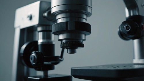
Have you ever wondered how doctors can operate on the most minor parts of our bodies? How can they manage such a small structure like a blood vessel in the brain, or heal the delicate nerves in a hand? The solution to this can be found in a spectacular device, a surgical microscope. This fantastic machine has transformed the mode of operation for doctors to the point where they can see and work on body parts that could not be mended at all. What is a Surgical Microscope? Imagine a magnifying glass with a superpower that is taken into the doctors ' hands when they operate. But it is not all about making things look bigger. A surgical microscope is a type of microscope used inside an operating room. It enhances the viewer by allowing them to see small structures, a close-up view of a small body part, such that surgeons can make out what the naked eye cannot see. In contrast to other microscopes commonly found in science classes, surgical microscopes are designed to operate while performing surgery in a standing position. They possess special lighting, many lenses, and they are easily movable during an operation. The microscope is placed on a stand that can be positioned at varying heights and angles to ensure comfort when the surgeon is using it. The Secret to the Magnification The primary role of a surgical microscope is to enlarge minor aspects. The magnification power of most surgical microscopes is up to 40 times the actual length of the object. In comparison, consider what would be seen when you put a grain of rice under one of these microscopes; it would look bigger than a marble, in fact, even larger than that. The system of magnification is achieved through the combination of lenses that work together like a team. The microscope features an objective lens to collect light in the surgical field, and there are eyepieces through which the surgeon views the specimen. Some current surgical microscopes even have cameras built into them, allowing the rest of the surgical team to view a screen displaying what the surgeon is looking at. Why Do Physicians Require Such Clear Vision? You may be confused as to why the doctors have to look into things closely when operating. The human body is a complex system, and even the smallest structures are crucial to our health. Blood vessels may be as narrow as a hair, nerves may be smaller than that of a thread, and there are parts in organs which are almost microscopic. When attacked by injury or disease, these minute structures must be repaired with the utmost precision. With a surgical microscope, it would be like attempting to fix a watch while wearing heavy mittens and sunglasses, which is almost impossible to do with precision, just as without. 1. Brain Surgery: Brain surgery is among the most advanced applications of surgical microscopes. The brain is a highly complicated composite of tiny blood vessels, neuron cells, and other cellular matter. When a surgeon has to remove a tumor or even repair his damaged blood vessels in his brain, he is expected to go very slowly so that he does not end up destroying healthy tissue around him. A surgical microscope enables brain surgeons to distinguish between normal and diseased tissue. They identify small blood vessels to leave without harming the regions that regulate speech, movements, or memory. This degree of precision has made brain surgery very safe and successful, much like it was in the past. 2. Eye Surgery: Eyes are one of the most sensitive parts of our body. The tiny structures within the eye are also delicate to the extent that any minor error during surgery can result in loss of sight. Eye surgery almost always requires surgical microscopes, whether for cataract or retinal procedures. In case of cataract surgery, the surgeons will be required to remove the clouded lens contained within the eye cavity and replace it with an artificial lens. This surgery is done through a few millimeters slit. The surgical microscope provides the surgeon with a clear view of the minute structures present in the eyes, enabling surgery to be performed with great precision. 3. Surgery of Hand and Nerve: If a person sustains a serious injury to the hand and fingers, they may damage small nerves and blood vessels that are essential for sensation and motion. Due to their size (which tends to be smaller than the tip of a pencil), repairing these structures needs the assistance of a surgical microscope. This is because, by using the microscope, the surgeon can meticulously rejoin cut nerves and blood vessels, a technique known as microsurgery. Such a surgery has the potential to restore the feeling and working abilities of injured hands and fingers, which would not be possible without the microscope-supplied enhanced vision. 4. Ear Surgery: The inner ear holds some of the tiniest bones in the human body, coupled with minute parts that undertake hearing and equilibrium. Surgical microscopes help doctors in fixing these small parts when they are broken. An example would be where the microscopic bones in the middle ear are worn out, the surgeon, with the aid of a microscope, would reconstruct them bit by bit or even substitute them with small artificial ones. This surgical procedure has the capability of restoring hearing to individuals who have lost it due to injury or illness. The Positive Side A lighting system is among the most valuable elements of a surgical microscope. This microscope features special lights that provide bright, even illumination to the surgical area. This lighting is essential, as magnifying an object also leads to darkening of the item, just as with a normal magnifying glass. A surgical microscope has intense lights that don't produce heat, preventing tissue damage. They also shed light in various directions, thereby causing no shadows, and the surgeon can notice every minute detail. 1. New Achievements: Surgical Microscopes, which are used today, are significantly superior compared to those used in the past. Most of them now have high-definition cameras that enable them to capture everything during the surgery. This will aid in educating other doctors and facilitate a review of the surgery at a later date. Special filters are also incorporated into some microscopes, providing surgeons with a clearer view of various tissues. Notably, some filters may cause blood vessels to appear larger, while others may facilitate the detection of diseased tissue. 2. The Human Touch: Training and skill Though surgical microscopes are unbelievable equipment, they are just as effective as the surgeons using them. It will take several years before a learner can operate a microscope effectively. Surgeons must develop a steady hand and train themselves to work with a small depth of field, as the image seen under the microscope is slightly flattened compared to natural sight. Some surgeons undergo several hundred hours of training on models and simulations of real-life scenarios before operating on their first patient using the microscope, thereby becoming proficient in microscopic surgery. The importance of this training lies in the fact that there is little room for error when handling extremely small objects. Tiny Surgery of The Future The surgical microscope is continually evolving as technology advances. Particularly, new models can offer computer support devices that direct the surgeon's movements, and others have a high imaging capacity, providing more detailed information about the area of operation. The use of surgical microscopes has significantly changed the working aspect of medicine, as body parts that were previously believed to be beyond repair can now be fixed. Such wonderful machines not only bring life to people and improve the lives of thousands of patients on this planet, but the small details also show that even the tiniest things can make a difference.
Slit Lamp vs. Other Ophthalmic Instruments: When to Use What?
In ophthalmology, accurate and precise diagnosis is crucial for effective treatment. The industry has seen the introduction of various instruments to assist eye care professionals in measuring different attributes of the eye. Although there are several instruments, the slit lamp is the mainstay. However, how does the slit lamp measure against other ocular instrumentation, and when should each be used? This blog will outline the differences between these instruments, describe their practical applications, and explain the nature of some basic eye care tools used in clinical settings. What is a Slit Lamp? Slit lamp - the specialized microscope in ocular medicine for examination of anterior and posterior segments-eye pathology under high and direct illumination of the slit beam. It consists of a light source adjustable to a narrow slit-in this case, the shine by the name, and its binocular microscope. When combined with these, additional lenses with different auxiliary apparatus, such as a tonometer or fundus lens, transform it into a powerful device for examining the entire eye, from the cornea to the retina. The examination will include findings about the conjunctiva, the cornea, the anterior chamber, the iris, the lens, and even the vitreous body. With those above special diagnostic lenses (90D or 78D), the slit lamp can also view the retina and optic nerve. Versatility and precision render it invaluable in routine eye examination and for diagnosing cataracts, corneal ulcers, glaucoma, and retinal detachments. Slit Lamp's Performance Compared to Other Instruments While the slit lamp is indeed flexible, it is by no means the only instrument that an ophthalmologist uses. Different instruments serve their purpose. Depending on the area of interest, a practitioner may turn to a variety of other instruments, such as a tonometer, autorefractor, ophthalmoscope, or an optical coherence tomography (OCT) device. Each instrument, therefore, provides unique data that contributes to a comprehensive understanding of the patient's ocular health. So, let's discuss every tool in more detail; then we can compare it with the slit lamp to know when to use it. 1. Slit Lamp Vs. Direct and Indirect Ophthalmoscope Among the oldest tools of the ophthalmologist, the direct ophthalmoscope is a handheld device that enables an enlarged view of the retina within a confined field of vision. However, this is so because it is portable and helpful in making rapid inspections, especially in emergency or field situations. In contrast to the clear view offered by an indirect ophthalmoscope, the perception of depth is significantly improved, as it provides a wider field of view, especially when examining peripheral retinal lesions or detachments. However, this technique requires pupil dilation and a relatively high level of skill to operate it effectively. As against these, the slit lamp—in conjunction with a condensing lens—offers a higher degree of magnification and a stereoscopic view of the retina, making it appropriate for referring examinations of the optic disc, macula, and blood vessels. While the other eyepieces will be helpful for general assessments and travel examinations, the slit lamp will be preferred over others for thorough all-in-one in-clinic prying. 2. Slit Lamp vs Tonometry Tonometry is the precise procedure for measuring intraocular pressure (IOP), a crucial parameter used to diagnose and monitor glaucoma conditions. The Goldmann applanation tonometer is considered the gold standard and is mounted on the slit lamp. It flattens a small area of the cornea to estimate IOP. There are other types of tonometers, such as non-contact (air puff) or rebound tonometers which are more user-friendly since they do not require a slit lamp as part of the setup. While tonometry provides a measure of a specific parameter, the slit lamp is used to determine the structural integrity of the angle for diseases such as narrow-angle glaucoma, inflammation, or trauma. Thus, tonometry is usually performed in conjunction with slit lamp examination rather than as a substitute for it. 3. Slit Lamp vs Autorefractor and Phoropter Autorefractors and phoropters are the preferred instruments for ascertaining refractive error. An autorefractor is an objective measurement of refractive status; however, a phoropter aids the optometrist in refining the prescription with subjective measurements. The slit lamp has no direct bearing on refractive error measurements and offers a strength that is the personnel's anatomical assessment of the eye. The slit lamp can aid in the recognition and documentation of cataracts or corneal pathology. If the patient's visual acuity is decreased, these are additional factors to consider about ocular health. 4. Slit Lamp vs OCT Optical Coherence Tomography (OCT) is another cutting-edge technique used to yield crystal-clear cross-sectional images of the retina, optic nerve, and the back portion of the eye. OCT is a highly effective imaging technique for diagnosing and monitoring a range of ocular issues, including macular degeneration, diabetic retinopathy, and glaucoma. The slit lamp, although a vital imaging tool, has its downsides, as it does not provide layer-by-layer assessment along with quantitative data, whereas OCT can do so easily. The slit lamp plays a crucial role in identifying visible structural changes that may warrant an OCT examination. When we use the slit lamp and OCT together in practice, we are looking for anatomical changes responsible for decreased visual acuity using the slit lamp, and then using the OCT to gain a deeper understanding of what we have observed. 5. Fundus Camera vs Slit Lamp A fundus camera is an instrument that captures color images of the eye's internal structures, such as the retina and optic nerve. This instrument is most effective in scenarios where tracking disease progression is a crucial component of the treatment. Capturing photographs can be done quickly and effortlessly using this camera, and the images can be sent to the specialists for secondary diagnosis. The fundus camera provides static images compared to the slit lamp, which offers the capability to observe the eye's surface in real-time with various lighting and magnification options. Both are necessary applications in modern ophthalmology —the slit lamp for documentation and the fundus camera for documentation and patient education. 6. Slit Lamp vs Corneal Topographer Corneal topography shows the point of curvature and shape of the cornea. The imaging of topography is beneficial in fitting contact lenses, identifying candidates for refractive surgery, and diagnosing keratoconus. It provides a detailed color-coded map that a slit lamp cannot do. Although the slit lamp excels at observing opacities and irregularities on the corneal surface, it cannot quantify and map the shape. Therefore, slit lamp corneal topography serves as a supporting imaging technique in surgical practice, providing precise and informative measurements. Both should be used together for the best results. Conclusion The slit lamp is a necessary part of ophthalmic diagnostics and provides a detailed and versatile way to examine the eye. But it is only part of the diagnostic equation. However, for a truly comprehensive approach to patient care, all instruments have a part to play, giving the eye professional a clear and complete picture of the issue. Considering these tools as substitutes is the wrong approach; instead, eye care professionals should view them as complementary to one another if they aim for a correct diagnosis and assessment. All of these tools are part of the eye doctor's toolkit and should be used correctly and at the right time to ascertain the patient's symptoms and history.
Beyond the Basics: A Layman's Guide to the Slit Lamp Microscope
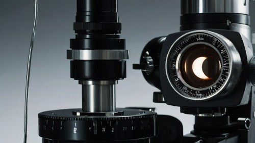
You have likely seen the intimidating machine with lights and mirrors that you sit in front of during an eye test with a doctor. The doctor puts their ear to the wall, tinkers with several knobs, and looks into a small telescope at your face. It is a slit lamp microscope, one of the most significant tools used in eye care. The little slit lamp may look elaborate, but it is a genius yet simple idea that assists doctors in examining the eye with fine detail. So, here is a more straightforward explanation of this fantastic piece of medical equipment. What Exactly Is a Slit Lamp Microscope? Consider a slit lamp as a high-powered magnifying glass and a very accurate, pointed flashlight. The slit component is the result of the small beam of light produced by it, which can be made narrow by a small line or widened into a thicker beam. The lamp provides the light, and the microscope section magnifies everything, allowing the doctor to see small details. The entire system is placed on a table, and both you and the doctor occupy specific positions. The doctor looks through the microscope section, and you put your chin on a rest, staring straight ahead. They are designed to provide the doctor with a clear view of your eyes while ensuring your comfort and well-being. It does the magic as the light beam falls on your eye at an angle, as the doctor's observation is made through the microscope at an angle. This arrangement provides a three-dimensional image of your eye structures, which would otherwise be impossible to observe. The Two Main Parts: Light and Magnification The Illumination System The light system is more similar to a super-precise flashlight, which can be regulated in numerous ways. A bright, clean, white light is provided by a bright halogen or LED bulb, usually used as a light source. Some filters and lenses reflect this light and end up in your eye. It is an adjustable slit. How about peeping through a keyhole compared to peeking through a door? The slit functions just like that. A thin beam can cut through various layers in your eye, and a wide slit lets in a wide range of light. The doctor is also able to adjust the position of the light, making it appear in various directions. This is particularly important since certain eye complications manifest only when light is directed to a specific side of the eye. The Observation System The microscope itself is a highly advanced pair of binoculars attached to an adjustable stand. It also gives a magnification of approximately 6-40 times the standard size, depending on the model and the settings. The number of different magnification levels that the doctor can adjust on most slit lamps is extensive. Lower magnification shows the entire eye area, while high magnification allows the doctor to focus on specific details. Designed as binoculars, it provides depth. The three-dimensional picture helps physicians determine the magnitude of another structure or how deeply issues may extend within the eye. The Process of Examination: The first time you sit down for a slit lamp exam, it may seem like a mysterious process, but it is pretty simple. The physician begins by aligning the equipment at your height level and eye position. Doctors examine your eyes, including your eyelashes and eyelids, as well as the cornea. They may request that you blink or look in various directions to indicate how everything moves. Different Types of Light Techniques There are several lighting methods applied in the examination by doctors, and each technique provides different information: · Direct illumination is similar to placing a flashlight on an object. This highlights surface-level information and apparent issues. · Indirect illumination is reflected against the structures around to produce subliminal effects of light. This method makes visible issues that are concealed in direct light. · Retro illumination features light that emanates from around the structures and glows. This is ideal for examining clear areas of the eye and detecting cloudiness, as well as any abnormalities. What Can Doctors View Using Such an Incredible Tool? The slit lamp provides a fantastic level of detail about your eyes. Doctors are capable of examining all major structures, both internally and externally. The Front of the Eye: The eyelids and the eyelashes may appear to be a simple thing, but they can contain numerous issues. The slit lamp can detect minute mites, oil glands blocked, or indications of inflammation that the naked eye cannot detect. The clear portion of your eye (called the sclera) is closely scrutinized, and the white one is also scrutinized, as well as the clear covering above the colored portion of your eye (conjunctiva). The color (redness) and swelling or abnormal development are clear under a microscope. The Cornea: The slit lamp excels most significantly in examining the cornea. This thick, dome-like anterior part of your eye is only half a millimeter thick, though the slit lamp may scan through this in layers. Doctors detect scratches, infections, and cloudy areas that blur vision. They measure cornea thickness and check for signs of swelling, which may indicate potential problems. In the Anterior Chamber: The back of the cornea contains clear fluid known as the anterior chamber. A slit lamp can examine this space to determine whether it is inflamed, bleeding, or otherwise. Your eye color (the iris) and the black hole in the center of the eye (the pupil) are closely checked as well. Discolorations of the iris or abnormal shapes of the pupil may indicate any eye disease. Why This Tool Is So Important? The slit lamp microscope is a marvel that has revolutionized eye care, as it enables doctors to detect any problem that would have remained unseen until it became severe. Most eye diseases begin with minor alterations that are not noticeable to the naked eye in normal lighting conditions. Many eye diseases should be identified early. Another example is glaucoma, which can gradually rob you of sight, with no signs until the damage is in an advanced stage. The slit lamp can help diagnose or detect the onset of glaucoma even before you are aware of it. Special Techniques and Add-ons Contemporary slit lamps are capable of much more than simple analysis. Most of them have special attachments and methods that vastly increase their capabilities. Pressure Measurement Certain slit lamps can even measure pressure in your eye (by applanation tonometry). A tiny probe is softly placed on the front of your eye (after numbing drops) to gauge the amount of pressure required to distribute the inside of a highly tiny region. The test plays a critical role in the identification of glaucoma. Documentation and Photography Many contemporary slit lamps are equipped with cameras, which can capture images of what the physician sees up close. Such images can be stored in your medical file, sent to other physicians, or used to track changes over time. Fluorescein Staining Physicians sometimes use fluorescein, an orange dye, to highlight specific eye issues. It shows scratches, dry areas, or injuries on the eye surface and appears bright green under the slit lamp's blue light. The Human Touch in High-Tech Medicine Despite all its high technology, slit lamp still needs human talent to become truly useful. It takes years for doctors to become trained in the use of all the various lighting techniques, as well as to understand what they observe. It is an art to know which technique to apply and when, how to vary the lighting according to the patient, and to identify specific nuances that may suggest serious complications. A computer cannot substitute a trained eye and the experience of an expert eye care specialist. Final Words This slit lamp microscope may appear to be a beast, but it is your weapon on the path to healthy eyes. This is a highly advanced piece of equipment that enables physicians to examine the eyes in great detail, detect early defects, and monitor treatment progress. The next time you are in front of one, you will be aware that you are using one of the most advanced and notable instruments of modern medicine.

