Blogs
Slit Lamp vs. Other Ophthalmic Instruments: When to Use What?

In ophthalmology, accurate and precise diagnosis is crucial for effective treatment. The industry has seen the introduction of various instruments to assist eye care professionals in measuring different attributes of the eye. Although there are several instruments, the slit lamp is the mainstay. However, how does the slit lamp measure against other ocular instrumentation, and when should each be used? This blog will outline the differences between these instruments, describe their practical applications, and explain the nature of some basic eye care tools used in clinical settings.
What is a Slit Lamp?
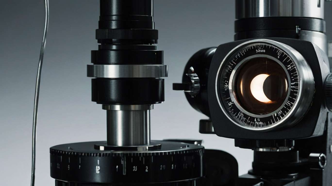
Slit lamp - the specialized microscope in ocular medicine for examination of anterior and posterior segments-eye pathology under high and direct illumination of the slit beam. It consists of a light source adjustable to a narrow slit-in this case, the shine by the name, and its binocular microscope. When combined with these, additional lenses with different auxiliary apparatus, such as a tonometer or fundus lens, transform it into a powerful device for examining the entire eye, from the cornea to the retina.
The examination will include findings about the conjunctiva, the cornea, the anterior chamber, the iris, the lens, and even the vitreous body. With those above special diagnostic lenses (90D or 78D), the slit lamp can also view the retina and optic nerve. Versatility and precision render it invaluable in routine eye examination and for diagnosing cataracts, corneal ulcers, glaucoma, and retinal detachments.
Slit Lamp's Performance Compared to Other Instruments
While the slit lamp is indeed flexible, it is by no means the only instrument that an ophthalmologist uses. Different instruments serve their purpose. Depending on the area of interest, a practitioner may turn to a variety of other instruments, such as a tonometer, autorefractor, ophthalmoscope, or an optical coherence tomography (OCT) device.
Each instrument, therefore, provides unique data that contributes to a comprehensive understanding of the patient's ocular health. So, let's discuss every tool in more detail; then we can compare it with the slit lamp to know when to use it.
1. Slit Lamp Vs. Direct and Indirect Ophthalmoscope
Among the oldest tools of the ophthalmologist, the direct ophthalmoscope is a handheld device that enables an enlarged view of the retina within a confined field of vision. However, this is so because it is portable and helpful in making rapid inspections, especially in emergency or field situations.
In contrast to the clear view offered by an indirect ophthalmoscope, the perception of depth is significantly improved, as it provides a wider field of view, especially when examining peripheral retinal lesions or detachments. However, this technique requires pupil dilation and a relatively high level of skill to operate it effectively.
As against these, the slit lamp—in conjunction with a condensing lens—offers a higher degree of magnification and a stereoscopic view of the retina, making it appropriate for referring examinations of the optic disc, macula, and blood vessels. While the other eyepieces will be helpful for general assessments and travel examinations, the slit lamp will be preferred over others for thorough all-in-one in-clinic prying.
2. Slit Lamp vs Tonometry
Tonometry is the precise procedure for measuring intraocular pressure (IOP), a crucial parameter used to diagnose and monitor glaucoma conditions. The Goldmann applanation tonometer is considered the gold standard and is mounted on the slit lamp. It flattens a small area of the cornea to estimate IOP.
There are other types of tonometers, such as non-contact (air puff) or rebound tonometers which are more user-friendly since they do not require a slit lamp as part of the setup. While tonometry provides a measure of a specific parameter, the slit lamp is used to determine the structural integrity of the angle for diseases such as narrow-angle glaucoma, inflammation, or trauma. Thus, tonometry is usually performed in conjunction with slit lamp examination rather than as a substitute for it.
3. Slit Lamp vs Autorefractor and Phoropter
Autorefractors and phoropters are the preferred instruments for ascertaining refractive error. An autorefractor is an objective measurement of refractive status; however, a phoropter aids the optometrist in refining the prescription with subjective measurements.
The slit lamp has no direct bearing on refractive error measurements and offers a strength that is the personnel's anatomical assessment of the eye. The slit lamp can aid in the recognition and documentation of cataracts or corneal pathology. If the patient's visual acuity is decreased, these are additional factors to consider about ocular health.
4. Slit Lamp vs OCT
Optical Coherence Tomography (OCT) is another cutting-edge technique used to yield crystal-clear cross-sectional images of the retina, optic nerve, and the back portion of the eye. OCT is a highly effective imaging technique for diagnosing and monitoring a range of ocular issues, including macular degeneration, diabetic retinopathy, and glaucoma. The slit lamp, although a vital imaging tool, has its downsides, as it does not provide layer-by-layer assessment along with quantitative data, whereas OCT can do so easily.
The slit lamp plays a crucial role in identifying visible structural changes that may warrant an OCT examination. When we use the slit lamp and OCT together in practice, we are looking for anatomical changes responsible for decreased visual acuity using the slit lamp, and then using the OCT to gain a deeper understanding of what we have observed.
5. Fundus Camera vs Slit Lamp
A fundus camera is an instrument that captures color images of the eye's internal structures, such as the retina and optic nerve. This instrument is most effective in scenarios where tracking disease progression is a crucial component of the treatment. Capturing photographs can be done quickly and effortlessly using this camera, and the images can be sent to the specialists for secondary diagnosis.
The fundus camera provides static images compared to the slit lamp, which offers the capability to observe the eye's surface in real-time with various lighting and magnification options. Both are necessary applications in modern ophthalmology —the slit lamp for documentation and the fundus camera for documentation and patient education.
6. Slit Lamp vs Corneal Topographer
Corneal topography shows the point of curvature and shape of the cornea. The imaging of topography is beneficial in fitting contact lenses, identifying candidates for refractive surgery, and diagnosing keratoconus. It provides a detailed color-coded map that a slit lamp cannot do.
Although the slit lamp excels at observing opacities and irregularities on the corneal surface, it cannot quantify and map the shape. Therefore, slit lamp corneal topography serves as a supporting imaging technique in surgical practice, providing precise and informative measurements. Both should be used together for the best results.
Conclusion
The slit lamp is a necessary part of ophthalmic diagnostics and provides a detailed and versatile way to examine the eye. But it is only part of the diagnostic equation. However, for a truly comprehensive approach to patient care, all instruments have a part to play, giving the eye professional a clear and complete picture of the issue. Considering these tools as substitutes is the wrong approach; instead, eye care professionals should view them as complementary to one another if they aim for a correct diagnosis and assessment. All of these tools are part of the eye doctor's toolkit and should be used correctly and at the right time to ascertain the patient's symptoms and history.
