Blogs
The Tiny Details That Matter: What is a Surgical Microscope and Why is it Used?

Have you ever wondered how doctors can operate on the most minor parts of our bodies? How can they manage such a small structure like a blood vessel in the brain, or heal the delicate nerves in a hand? The solution to this can be found in a spectacular device, a surgical microscope.
This fantastic machine has transformed the mode of operation for doctors to the point where they can see and work on body parts that could not be mended at all.
What is a Surgical Microscope?
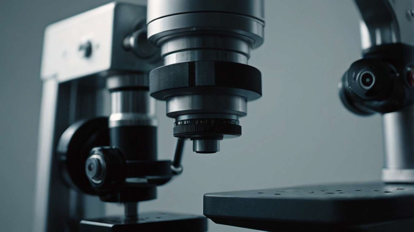
Imagine a magnifying glass with a superpower that is taken into the doctors ' hands when they operate. But it is not all about making things look bigger. A surgical microscope is a type of microscope used inside an operating room. It enhances the viewer by allowing them to see small structures, a close-up view of a small body part, such that surgeons can make out what the naked eye cannot see.
In contrast to other microscopes commonly found in science classes, surgical microscopes are designed to operate while performing surgery in a standing position. They possess special lighting, many lenses, and they are easily movable during an operation. The microscope is placed on a stand that can be positioned at varying heights and angles to ensure comfort when the surgeon is using it.
The Secret to the Magnification
The primary role of a surgical microscope is to enlarge minor aspects. The magnification power of most surgical microscopes is up to 40 times the actual length of the object. In comparison, consider what would be seen when you put a grain of rice under one of these microscopes; it would look bigger than a marble, in fact, even larger than that.
The system of magnification is achieved through the combination of lenses that work together like a team. The microscope features an objective lens to collect light in the surgical field, and there are eyepieces through which the surgeon views the specimen. Some current surgical microscopes even have cameras built into them, allowing the rest of the surgical team to view a screen displaying what the surgeon is looking at.
Why Do Physicians Require Such Clear Vision?
You may be confused as to why the doctors have to look into things closely when operating. The human body is a complex system, and even the smallest structures are crucial to our health. Blood vessels may be as narrow as a hair, nerves may be smaller than that of a thread, and there are parts in organs which are almost microscopic.
When attacked by injury or disease, these minute structures must be repaired with the utmost precision. With a surgical microscope, it would be like attempting to fix a watch while wearing heavy mittens and sunglasses, which is almost impossible to do with precision, just as without.
1. Brain Surgery:
Brain surgery is among the most advanced applications of surgical microscopes. The brain is a highly complicated composite of tiny blood vessels, neuron cells, and other cellular matter. When a surgeon has to remove a tumor or even repair his damaged blood vessels in his brain, he is expected to go very slowly so that he does not end up destroying healthy tissue around him.
A surgical microscope enables brain surgeons to distinguish between normal and diseased tissue. They identify small blood vessels to leave without harming the regions that regulate speech, movements, or memory. This degree of precision has made brain surgery very safe and successful, much like it was in the past.
2. Eye Surgery:
Eyes are one of the most sensitive parts of our body. The tiny structures within the eye are also delicate to the extent that any minor error during surgery can result in loss of sight. Eye surgery almost always requires surgical microscopes, whether for cataract or retinal procedures.
In case of cataract surgery, the surgeons will be required to remove the clouded lens contained within the eye cavity and replace it with an artificial lens. This surgery is done through a few millimeters slit. The surgical microscope provides the surgeon with a clear view of the minute structures present in the eyes, enabling surgery to be performed with great precision.
3. Surgery of Hand and Nerve:
If a person sustains a serious injury to the hand and fingers, they may damage small nerves and blood vessels that are essential for sensation and motion. Due to their size (which tends to be smaller than the tip of a pencil), repairing these structures needs the assistance of a surgical microscope.
This is because, by using the microscope, the surgeon can meticulously rejoin cut nerves and blood vessels, a technique known as microsurgery. Such a surgery has the potential to restore the feeling and working abilities of injured hands and fingers, which would not be possible without the microscope-supplied enhanced vision.
4. Ear Surgery:
The inner ear holds some of the tiniest bones in the human body, coupled with minute parts that undertake hearing and equilibrium. Surgical microscopes help doctors in fixing these small parts when they are broken.
An example would be where the microscopic bones in the middle ear are worn out, the surgeon, with the aid of a microscope, would reconstruct them bit by bit or even substitute them with small artificial ones. This surgical procedure has the capability of restoring hearing to individuals who have lost it due to injury or illness.
The Positive Side
A lighting system is among the most valuable elements of a surgical microscope. This microscope features special lights that provide bright, even illumination to the surgical area. This lighting is essential, as magnifying an object also leads to darkening of the item, just as with a normal magnifying glass.
A surgical microscope has intense lights that don't produce heat, preventing tissue damage. They also shed light in various directions, thereby causing no shadows, and the surgeon can notice every minute detail.
1. New Achievements:
Surgical Microscopes, which are used today, are significantly superior compared to those used in the past. Most of them now have high-definition cameras that enable them to capture everything during the surgery. This will aid in educating other doctors and facilitate a review of the surgery at a later date.
Special filters are also incorporated into some microscopes, providing surgeons with a clearer view of various tissues. Notably, some filters may cause blood vessels to appear larger, while others may facilitate the detection of diseased tissue.
2. The Human Touch: Training and skill
Though surgical microscopes are unbelievable equipment, they are just as effective as the surgeons using them. It will take several years before a learner can operate a microscope effectively. Surgeons must develop a steady hand and train themselves to work with a small depth of field, as the image seen under the microscope is slightly flattened compared to natural sight.
Some surgeons undergo several hundred hours of training on models and simulations of real-life scenarios before operating on their first patient using the microscope, thereby becoming proficient in microscopic surgery. The importance of this training lies in the fact that there is little room for error when handling extremely small objects.
Tiny Surgery of The Future
The surgical microscope is continually evolving as technology advances. Particularly, new models can offer computer support devices that direct the surgeon's movements, and others have a high imaging capacity, providing more detailed information about the area of operation.
The use of surgical microscopes has significantly changed the working aspect of medicine, as body parts that were previously believed to be beyond repair can now be fixed. Such wonderful machines not only bring life to people and improve the lives of thousands of patients on this planet, but the small details also show that even the tiniest things can make a difference.
